Media

Instagram post

@macromoltek

Nov. 27, 2017

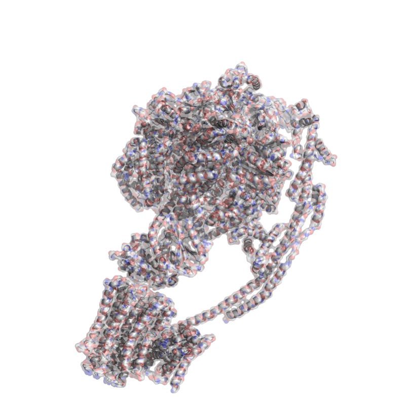
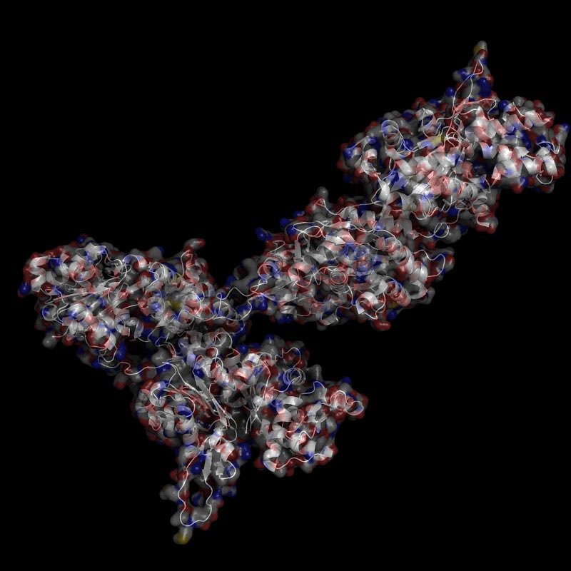
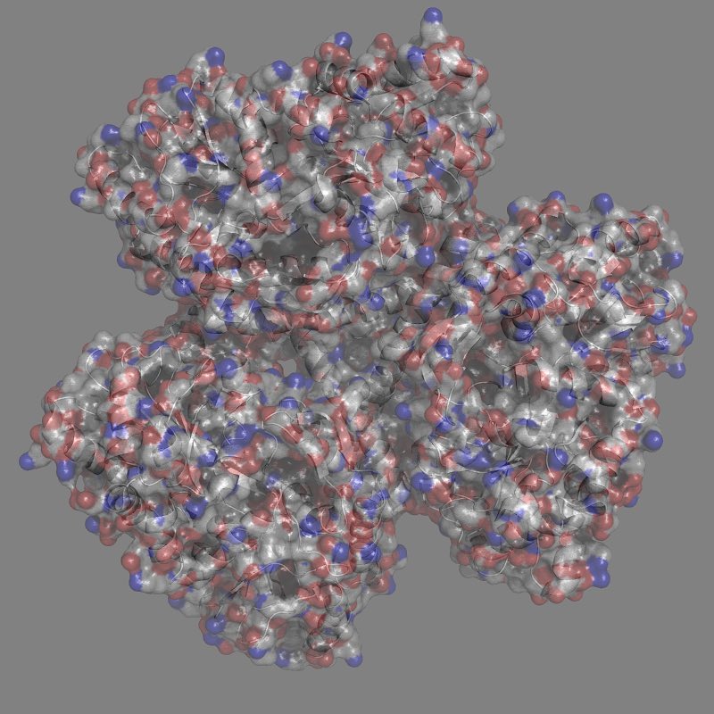
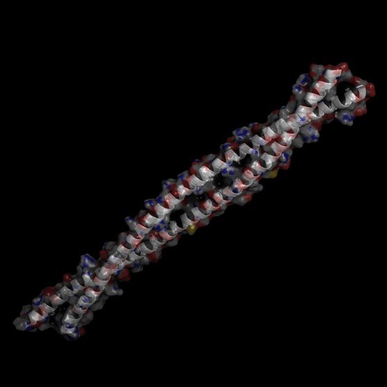
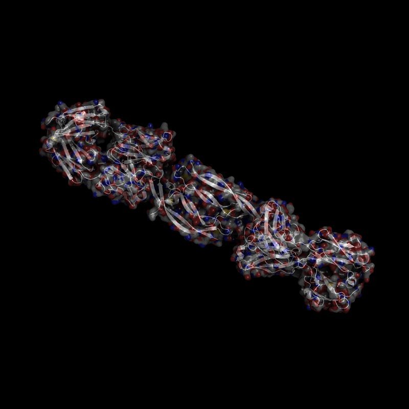
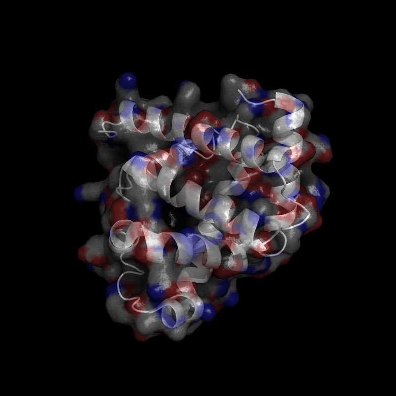
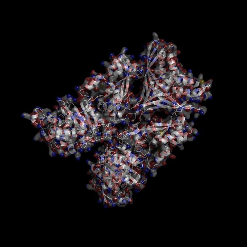
Our immune systems include a long list of defenses ready to stop pathogens every step of the way. The very first of these defenses is the outer layer of our skin called the epidermis. The epidermis is made of specialized cells called keratinocytes specifically named because they produce a structural protein called keratin en mass. The life of keratinocytes is really quite interesting! Keratinocytes start their lives as epidermal stem cells and randomly become a short-lived progenitor called a “transit amplifying cell”. This cell type continues to divide and migrate towards the outer layer of the epidermis. Along the way, the cell differentiates a few times and takes on new behaviors and characteristics. The final stage of the keratinocyte is the corneocyte. This cell is characterized by exiting the cell cycle, losing its nucleus and cytoplasmic organelles, and having its plasma membrane replaced with a host of keratin-proteins. This new keratin membrane is enveloped by an insoluble amalgam of proteins and linked by transglutamines and lipids. That’s the skin you see and touch! It’s made of proteins and fats. In fact, keratin forms a whole list of structures including hair, nails, hooves, feathers, horns, etc. which distinguish themselves by amount and type of keratin produced. There are dozens of different types of keratin, but each are structurally quite similar as you can see from the picture which includes keratins 1 and 10. Info about differentiation from: https://www.proteinatlas.org/humanproteome/skin

Instagram post

@macromoltek

Oct. 16, 2017

Penicillin is a broad-spectrum antibacterial drug, but few of us have considered how this drug performs its primary function. Essentially, penicillin reduces the integrity of bacterial cell walls. Bacteria are constantly replenishing their cell walls when growing or reproducing much like we regenerate our skin. Expansion is accomplished via a network of enzymes and peptidoglycans, the cell wall “bricks”. Penicillin-binding proteins (PBP) belong to a subgroup of enzymes called transpeptidases and are the essential “brick-layers” for bacteria. PBPs catalyze the reaction between peptidoglycans by mediating the removal of D-alanine from the precursor of peptidoglycan. This results in a conformational change and binding of adjacent peptidoglycans. Penicillin acts as an inhibitor for this reaction. The structure of penicillin is very similar to that of the peptidoglycans, so PBP forms a bond with penicillin at the active site. In fact, it’s a covalent bond, so it’s irreversible! PBP is thence rendered inactive, peptidoglycans are left unlinked, and the bacterium’s cell wall is full of holes. Osmotic pressure builds on the bacterium as the cell wall further degrades and eventually leads to cell death. Better them than you! PBPs come in a variety. Pictured here is Penicillin-binding Protein 2a from a resistant strain of Staphylococcus Aureus strain 27r. This bacteria evolved a mutation which allows its PBP2a to resist binding to penicillin. PDB: 1VQQ